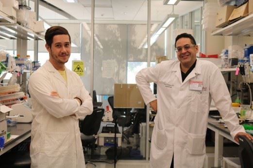
Gabriel Velez1,2,3 and Vinit B. Mahajan1,2,4
1Omics Laboratory, Stanford University, Palo Alto, CA
2Department of Ophthalmology, Byers Eye Institute, Stanford University, Palo Alto, CA
3Medical Scientist Training Program, University of Iowa, Iowa City, IA
4Veterans Affairs Palo Alto Health Care System, Palo Alto, CA
Precision Medicine aims to tailor medical therapies to individual patients by taking into account their specific genetics, environments, and lifestyle choices. Recently empowered by large sets of molecular and clinical data and high-powered analytics, this concept is changing the field of medicine, such that therapies can be customized for each patient [1]. Despite these advancements, the diagnosis and treatment of organ-specific inflammatory diseases, such as intraocular inflammation (i.e., uveitis), is most often empirical, relying heavily on clinical examination.
The inheritance and susceptibility to many inflammatory eye disorders is often polygenic and poorly understood [2]. Many rare genetic variants cannot be easily detected when screening the over 10,000 genes in the human genome. Thus, many of the recently developed genomic testing methods fail to pinpoint precise therapies for patients affected by these disorders, leaving many of them with limited treatment options. Genes are static: they do little to inform physicians when a disease will be active, when it will start, or when it will stop. For real-time analysis of a patient’s disease state, the attention should be directed towards proteins. Proteomic analysis is becoming an attractive and powerful method for characterizing the molecular profiles of diseased tissues, like the eye. Ophthalmologists have the ability to collect surgical tissue and fluid biopsies from patients and perform detailed molecular analysis.
To streamline the personalized proteomics pipeline for eye disease, our group has designed and implemented the Stanford Biorepository for Eye and Surgical Tissue (BEST), a novel system that allows for immediate and point-of-care processing of ophthalmic surgical samples. The system has several key components: a mobile operating-room cart with a flat lab-bench surface, a computer with secure access to a sample database, a barcode scanner, and lab supplies for sample processing away from the sterile surgical field [3]. Once biopsies are collected in the operating room, they are handed to a clinical research coordinator who processes the sample and transfers it to a 2D-barcoded cryotube for immediate flash-freezing. The barcode and corresponding patient phenotypic data is entered into the electronic database, allowing for efficient sample retrieval for downstream proteomic analyses [3, 4].
Once surgical specimens are properly collected and stored, their proteomic content can be analyzed using a variety of analytical platforms (Figure 1). Since human eye fluid (e.g., vitreous and aqueous humor) can contain several thousand unique proteins, liquid chromatography-tandem mass spectrometry (LC-MS/MS) and targeted proteomic platforms (e.g., multiplex ELISA arrays) should be used concurrently in the biomarker discovery phase to maximize the number of candidate biomarkers for prospective verification and validation studies [4, 5]. The use of these analytical platforms is exemplified in our study of the vitreous proteome from patients with Neovascular Inflammatory Vitreoretinopathy (NIV), a rare inherited inflammatory eye disease. Despite knowledge of the causative gene mutation underlying NIV (i.e., mutations in the CAPN5 gene), these patients were previously treated with non-specific immunosuppressive medications, such as oral corticosteroids and infliximab (anti-TNF-α). Characterization of the NIV vitreous proteome revealed that levels of TNF-α were indistinguishable from control vitreous, explaining the previous failure of infliximab therapy. Instead, we detected elevated levels of VEGF and IL-6. We therefore repositioned bevacizumab (anti-VEGF) and tocilizumab (anti-IL-6), which successfully halted neovascularization and cleared vitreous hemorrhages, reduced inflammatory cell infiltration, and mitigated intraocular fibrosis in these patients [6]. We have started to expand this investigative approach to include more common causes of blindness, such as retinitis pigmentosa, diabetic retinopathy, proliferative vitreoretinopathy, age-related macular degeneration, intraocular tumors, and uveitis [6-11].
Treatment of eye disease is now entering an era of Molecular Surgery [4]. In ophthalmology, routine microsurgical techniques can be used to safely extract fluid and tissue biopsies for downstream molecular analysis. Once molecular targets are identified, the same surgical techniques can be used to deliver precise molecular therapies in the operating room. Some of the most widely used drugs in ophthalmology today are biologics (e.g., anti-vascular endothelial growth factor [VEGF] antibodies) and engineered small molecules. It is common now to directly inject antibodies into the eye that bind to a specific protein target. As a result, physicians are becoming more aware of the exact molecular targets of the drugs they are giving patients. The same is true for a variety of immune diseases and cancers. Recasting eye surgery in molecular terms will allow scientists and ophthalmologists to take innovative approaches to curing blindness.
.png)
Figure 1. Personalized proteomics pipeline for precision medicine in ophthalmology: Liquid vitreous biopsies can be obtained in the operating room using a vitreous cutter or 23-gauge needle (left). Vitreous samples can be analyzed for protein content using multiplex ELISA arrays (top row). Custom or commercial antibody arrays quantify protein levels in biological samples using fluorescence or chemiluminescence means. Alternatively, vitreous fluid can be analyzed using a mass spectrometry approach (bottom row). Protein mixtures are digested with trypsin and peptides are extracted. Chromatography is used to separate peptides prior to ionization and mass acquisition by mass spectrometry. protein levels are quantified, downstream bioinformatics analysis (right) can help put the identified proteins into the context of the disease. Figure adapted from Velez, G., et al., Personalized Proteomics for Precision Health: Identifying Biomarkers of Vitreoretinal Disease. Transl Vis Sci Technol, 2018. 7(5): p. 12.
Bio
Dr. Gabriel Velez is a student and Alfred P. Sloan scholar in the Medical Scientist (MD/PhD) Training Program at the University of Iowa. He completed his PhD in Dr. Vinit B. Mahajan’s lab at the Byers Eye Institute at Stanford University where he used proteomic and structural biology methods to explore the mechanisms underlying inflammatory retinal diseases and identify therapeutic biomarkers. He is funded by an F30 training grant from the National Eye Institute and is currently finishing his final years of medical school at the University of Iowa. He plans to pursue residency training in ophthalmology after obtaining his MD degree.

References
1. Velez, G., et al., Personalized Proteomics for Precision Health: Identifying Biomarkers of Vitreoretinal Disease. Transl Vis Sci Technol, 2018. 7(5): p. 12.
2. Li, A.S., et al., Whole-Exome Sequencing of Patients With Posterior Segment Uveitis. Am J Ophthalmol, 2021. 221: p. 246-259.
3. Skeie, J.M., et al., A biorepository for ophthalmic surgical specimens. Proteomics Clin Appl, 2014. 8(3-4): p. 209-17.
4. Velez, G. and V.B. Mahajan, Molecular Surgery: Proteomics of a Rare Genetic Disease Gives Insight into Common Causes of Blindness. iScience, 2020. 23(11): p. 101667.
5. Skeie, J.M., C.N. Roybal, and V.B. Mahajan, Proteomic insight into the molecular function of the vitreous. PLoS One, 2015. 10(5): p. e0127567.
6. Velez, G., et al., Therapeutic drug repositioning using personalized proteomics of liquid biopsies. JCI Insight, 2017. 2(24).
7. Roybal, C.N., et al., Personalized Proteomics in Proliferative Vitreoretinopathy Implicate Hematopoietic Cell Recruitment and mTOR as a Therapeutic Target. Am J Ophthalmol, 2018. 186: p. 152-163.
8. Sepah, Y.J., et al., Proteomic analysis of intermediate uveitis suggests myeloid cell recruitment and implicates IL-23 as a therapeutic target. Am J Ophthalmol Case Rep, 2020. 18: p. 100646.
9. Velez, G., et al., Precision Medicine: Personalized Proteomics for the Diagnosis and Treatment of Idiopathic Inflammatory Disease. JAMA Ophthalmol, 2016. 134(4): p. 444-8.
10. Velez, G., et al., Proteomic insight into the pathogenesis of CAPN5-vitreoretinopathy. Sci Rep, 2019. 9(1): p. 7608.
11. Wert, K.J., et al., Metabolite therapy guided by liquid biopsy proteomics delays retinal neurodegeneration. EBioMedicine, 2020. 52: p. 102636.


.png)